
A Patient’s Guide to DynaROM Testing
The short version
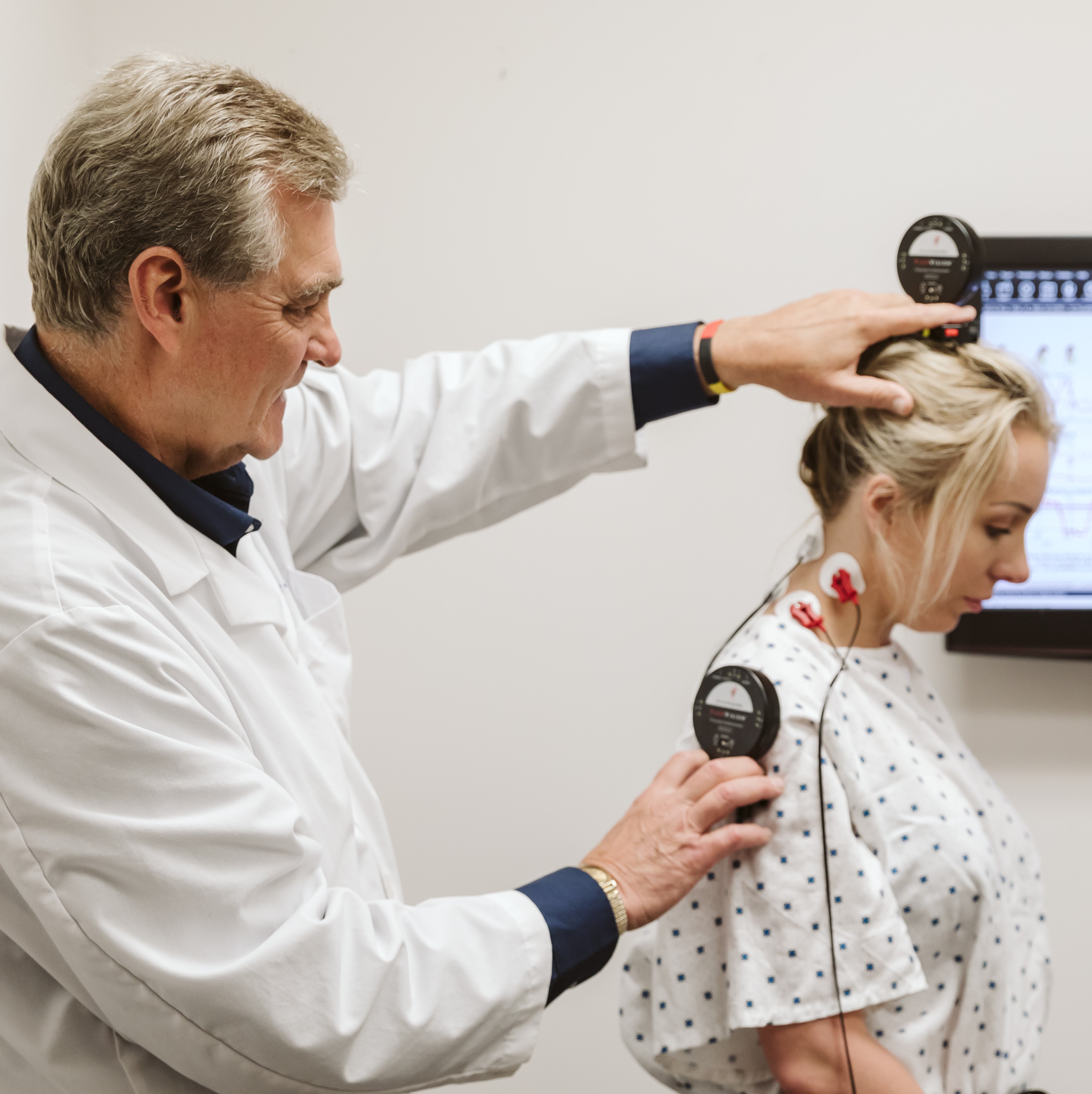 A Spinal EKG is the plain-language name for a test called DynaROM. It records the electrical activity in the muscles along your spine while you move, and at the same moment measures how far you move. It takes about 20 to 30 minutes, uses stick-on electrodes and two small wireless sensors, and does not hurt.
Its purpose is to answer a question that x-rays and MRIs cannot: what are your muscles actually doing when you bend and turn, and are they functioning normally or abnormally following an injury to that region? After a car crash, that question turns out to matter a great deal.
Why it is called a Spinal EKG
An electrocardiogram, the EKG you have probably had at a physical, records the electrical signal the heart muscle produces when it contracts, through electrodes placed on the skin. Every muscle in the body produces a signal like that. The technique of recording it from skeletal muscle through skin electrodes is called surface electromyography, usually shortened to surface EMG or sEMG.
The Spinal EKG applies that principle to the muscles that run along the spine. The name is a nickname rather than a formal one. The device is called the DynaROM, short for dynamic range of motion, and it is made by a Seattle company called MyoVision. The formal description of what it does is dynamic surface electromyography with simultaneous range of motion measurement. Spinal EKG is easier to say, and it captures the idea well enough: the same kind of recording, pointed at a different set of muscles.
What it actually measures
Two things at once, which is the whole point.
The first is electrical activity in the muscles, measured in microvolts through electrodes placed on the skin over the spinal muscles. This appears on the upper half of the results graph. The second is range of motion, measured in degrees by two small wireless sensors called inclinometers. This appears on the lower half. Because both are recorded at the same time, the graph shows not just how far you moved, but how much electrical activity your muscles were producing at every point along the way.
The word dynamic is doing important work in the name. Nearly every other test you have had for your spine was taken while you held still. This one is taken while you move, which is when most people with a soft tissue injury actually have symptoms.
What the test is looking for is a pattern called muscle guarding, the involuntary tightening of muscles around an injured joint to protect it. We explained guarding in detail in an earlier article on spasm, tightness, and stiffness after a crash. The short version is that guarding is a reflex. You cannot produce it on purpose and you cannot switch it off on purpose. On a Spinal EKG it shows up as muscles that stay active when they should be quiet, activity that spikes at a specific point in a movement, or one side of the spine working much harder than the other.
The one pattern worth understanding
There is a single graph pattern that explains most of what this test is for, and it is worth two minutes to understand.
When a healthy person bends forward as far as they can, the muscles of the lower back go quiet at the bottom of the bend. The weight of the upper body is being carried by ligaments and connective tissue at that point, and the muscles have nothing to do, so they relax. This is called the flexion-relaxation response, and it is one of the most consistent findings in spine physiology.
When the tissue in that region is injured, the muscles refuse to let go. They keep firing through the bottom of the bend, holding the spinal segment rigid to protect it. On the graph, a healthy result shows the muscle activity line dropping to near silence at full flexion. A guarded result shows it staying elevated, sometimes spiking, right where it should have gone quiet.
What the test is like
 Small adhesive electrodes are placed on the skin over the muscles being tested: for the neck, over the paraspinal muscles on both sides and the sternocleidomastoid muscles at the front; for the lower back, over the paraspinal muscles on both sides at the top and bottom of the lumbar spine. The wireless motion sensors are attached with a strap or headband, or held in place by the clinician. Then you are asked to move.
The standard protocol tests six motions: bending forward, bending backward, bending to each side, and turning to each side. You practice each motion first, then perform it two or three times over about thirty seconds while the system records. The recording itself takes only a few minutes; with setup, practice trials, and the review of results, the appointment generally takes about 20 to 30 minutes. Your range on each motion is reported in degrees and compared with the reference values published in the AMA Guides.
It is painless. There are no needles, no radiation, and nothing goes inside the body. You will need skin access at the neck or lower back, so a shirt that can be lifted or a gown works. Results appear on the screen as the test runs, a written report follows, and Dr. Schmidt reviews and interprets the findings with you after the test.
At this clinic the Spinal EKG is generally performed after the motion x-rays have been taken and read by a radiologist, so that the muscle findings can be interpreted alongside the ligament findings rather than on their own.
What normal and abnormal look like
A normal result has three features. The muscle activity is symmetric, so the left and right sides behave similarly on the same motion. The muscles quiet down where they are supposed to, particularly at full forward bend. And the range of motion curve is smooth, rising and falling without hitches.
An abnormal result usually breaks one or more of those. The two sides diverge. The muscles stay active where they should relax, or spike sharply at one point in the range. Or the motion curve itself looks jagged, with small stops and restarts along the way, sometimes described as ratcheting, which is what it looks like when a person moves through pain in increments.
Each of the six motions is graded separately by how abnormal the pattern is, and the report summarizes those grades. We cover how that grading works, and how to read it, in a companion article on how a Spinal EKG grades a soft tissue injury.
Why it is used after a car accident
We have written elsewhere about why the standard post-crash workup can come back normal while a real injury remains: x-rays, CT, and MRI are all still images of a spine at rest, and most soft tissue injury is a problem of motion. If that idea is new to you, the articles on spinal ligament injury and on a normal MRI with ongoing pain explain it fully.
The Spinal EKG fills a specific gap in that picture. Motion x-rays with computer measurement show how far the bones move relative to one another, which is the ligament question. The Spinal EKG shows how the muscles are responding to whatever is happening in that region, which is the guarding question. They are different questions, and a thorough soft tissue evaluation asks both.
There is a second reason the test matters, and it is best said plainly. Range of motion can be limited on purpose. A person can simply choose not to bend as far. Muscle guarding cannot be produced or suppressed on purpose, because it is a reflex. The American Medical Association’s Practical Guide to Range of Motion Assessment, which includes the DynaROM as a figure, makes exactly this point: recording muscle guarding alongside range of motion helps establish both true soft tissue injury and effort, because guarding cannot be feigned while range of motion can.
For a patient who has been told their symptoms are subjective, that is a meaningful thing. It is one of the few tests in this area whose result does not depend on what you say or how hard you try.
Tracking change over a course of care
A single Spinal EKG shows what your muscles are doing on the day it is performed. Its larger value comes from repeating it.
Testing at the start of care and again at intervals shows whether the guarding response is resolving, staying the same, or getting worse. That trajectory informs treatment decisions along the way, and it is one of the pieces of information a clinician uses in judging when a patient has reached maximum medical improvement, the point at which the condition has stabilized. The test does not predict how you will feel in the future. It documents how the muscles are behaving now and how that compares with the last testing, which informs whether the treatment plan continues as planned, changes, or calls for collaborative care planning with other medical professionals.
Where the test comes from
The DynaROM was developed by David Marcarian, a research scientist who trained and worked at NASA before founding MyoVision. The approach of recording range of motion and muscle activity simultaneously is patented (US Patent 9,808,172), and the patent describes the system as a soft-tissue-injury diagnostic system. A patent establishes that a method is novel and specifically defined; the clinical evidence behind it is a separate question, addressed below. The MyoVision system that performs the test is an FDA-cleared Class II medical device (510(k) K123399, cleared August 1, 2013, under the regulation for diagnostic electromyographs); the clearance covers providing the clinician with muscle electrical activity and motion-angle information, and it speaks to the device, not to any particular diagnostic conclusion drawn from it. Dr. Schmidt is certified by MyoVision in DynaROM testing.
The clearest outside endorsement comes from John Gerhardt, MD, the primary author of the American Medical Association’s Practical Guide to Range of Motion Assessment and himself the inventor of an earlier range of motion instrument. In a signed letter dated May 30, 2009, Dr. Gerhardt wrote that after evaluating the system on patients for nine years he had included it in the AMA text at page 45, Figure 2-27, and that the DynaROM had replaced the device he invented as, in his words, the new gold standard. The figure and its description appear in the current printing of that text.
Dynamic surface EMG also has its own procedure codes in the AMA’s Current Procedural Terminology system, CPT 96002 for the test and 96004 for its interpretation. To be assigned a Category I code, a procedure has to be consistent with contemporary medical practice, performed by many practitioners across the country, and supported by clinical efficacy documented in peer-reviewed literature. Dynamic surface EMG met those criteria. A code is not the same thing as insurance coverage, which is covered below.
What it cannot do, and the debate around it
It is worth being just as clear about the limits.
- It does not image anything. It cannot show a ligament, a disc, or a bone. Stress x-rays with computer measurement do that job, and a Spinal EKG does not replace them.
- It is not a nerve test. The EMG a neurologist orders, done with fine needles, evaluates nerve damage. That is a different test with a different purpose, and one does not substitute for the other.
- It is not universally accepted. Surface EMG has been debated in the medical literature for years. Several major insurers currently classify dynamic surface EMG as investigational when used to diagnose back or neck pain, and professional bodies have published mixed assessments. That debate is real, and we address it directly in a separate article rather than pretending it does not exist.
Our position is the one that fits the evidence. The Spinal EKG documents how the spinal muscles are behaving during motion, and it does so in a way that does not depend on the patient’s effort or account. It is a useful part of a thorough evaluation and a good way to track change over a course of care. It is not, on its own, proof of injury, and any clinic that presents it that way is overstating it. The manufacturer’s own report template says the same thing in its limitations section: no single test independently establishes injury or disability, and the results are to be interpreted together with the history, examination, imaging, and other clinical evidence.
Cost and insurance
The test is billed under AMA CPT codes 96002 and 96004. Coverage varies widely by insurer and some carriers do not cover it. Auto injury claims in Minnesota are covered under the No Fault Law; however, a minority of carriers may push back on claims. The most reliable answer will come from the clinic’s office manager who has verified benefits with the insurer.
When the emergency department comes first
A few symptoms after a crash need an emergency department rather than a clinic, and it is worth being clear about them. If you have a headache that is getting worse, weakness or numbness in an arm or leg, repeated vomiting, confusion, or any change in consciousness, go to an emergency department to rule out a life-threatening emergency.
For everyone else, which is most people after a crash, the evaluation begins at the clinic with a history, a physical and neurological examination, and imaging where it is indicated. Neurological findings such as weakness, numbness, or changed reflexes call for imaging, and where indicated medical referral, before any hands-on treatment. The Spinal EKG is part of evaluating the soft tissue injury that remains once a life-threatening emergency has been ruled out.
CONTACT US »
Frequently asked questions
Is a Spinal EKG the same as the EMG my neurologist ordered?
Is the Spinal EKG covered by insurance?
Who does DynaROM or Spinal EKG testing in Minnesota?